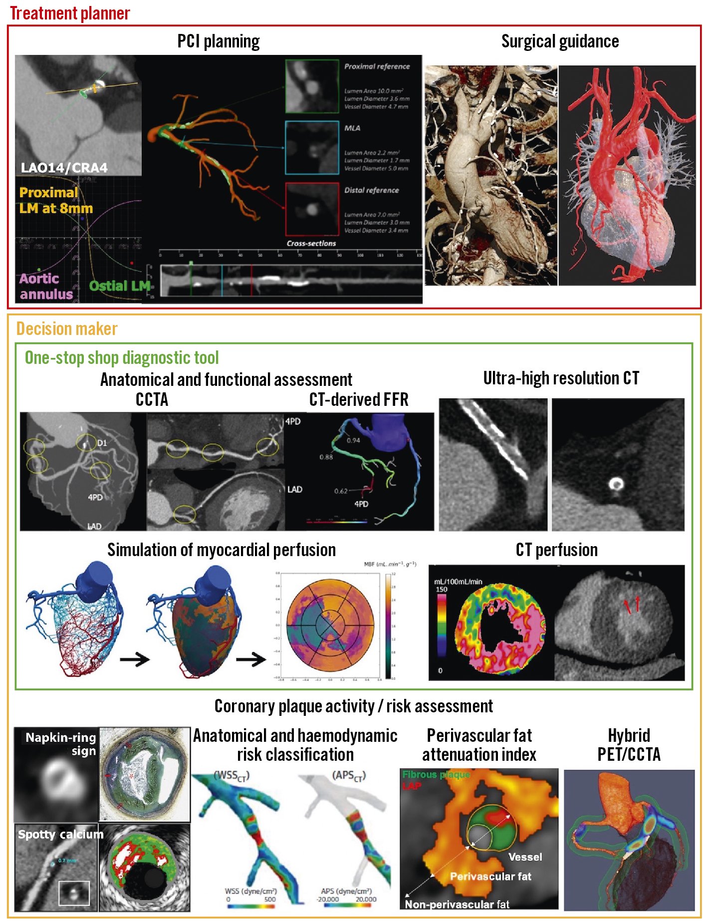
Angiography image processing tools enhance the visualization of blood vessels. They assist in accurate diagnosis and treatment planning.
Angiography is a critical medical imaging technique used to visualize the interior of blood vessels and organs. Advanced image processing tools are essential for improving the clarity and detail of angiographic images. These tools utilize sophisticated algorithms to highlight important features and remove noise, allowing healthcare professionals to make more accurate diagnoses.
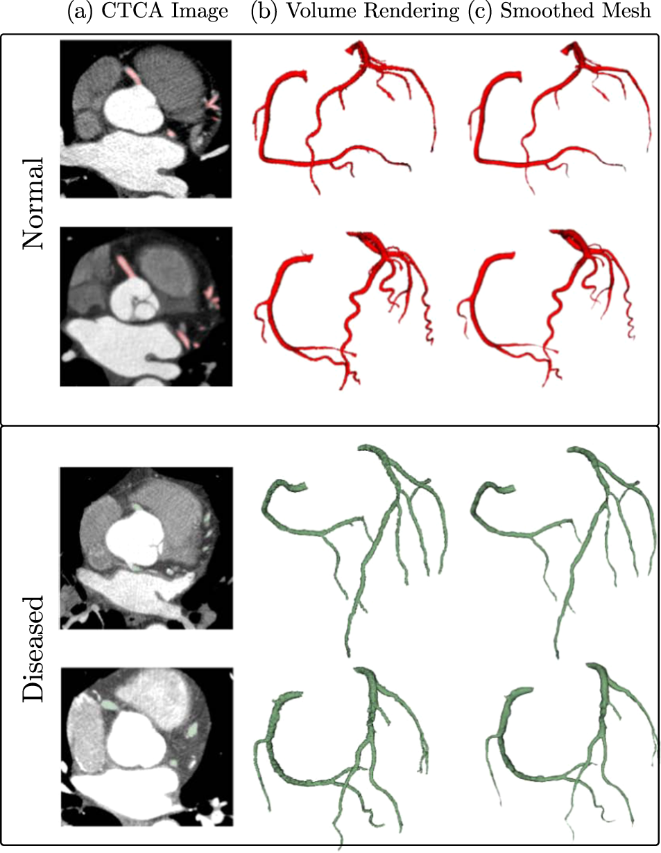
Enhanced visualization aids in the precise detection of abnormalities such as blockages or aneurysms. Modern angiography image processing tools integrate seamlessly with existing medical imaging systems, providing real-time analysis and 3D reconstruction capabilities. This ensures a higher level of patient care and optimized treatment strategies.
Introduction To Angiography
Angiography is a medical imaging technique. It helps visualize blood vessels. Doctors use it to diagnose and treat vascular conditions.
The Role Of Imaging In Cardiology
Imaging is crucial in cardiology. It provides clear pictures of the heart. These images help in detecting heart diseases. Doctors can see blockages and other issues. Imaging guides treatment plans effectively.
Evolution Of Angiography Techniques
Angiography techniques have evolved over time. Early methods were simple and less precise. Modern techniques are highly advanced. They provide detailed images with less risk.
| Old Techniques | Modern Techniques |
|---|---|
| Basic X-rays | CT Angiography |
| Invasive | Non-Invasive |
| Low Image Quality | High Image Quality |
- Early angiography was invasive.
- Modern techniques are safer.
- Image quality has greatly improved.
Angiography image processing tools are essential. They enhance the quality of images. These tools help in accurate diagnosis.
- Capture the image.
- Process the image.
- Analyze the image.
Understanding angiography is key. It helps in better heart disease management. Stay informed about the latest tools and techniques.
Key Angiography Modalities
Angiography is a medical imaging technique used to visualize blood vessels. Different modalities help in detecting issues in the vascular system. Each modality offers unique advantages and limitations.
Traditional X-ray Angiography
Traditional X-Ray Angiography uses X-rays to capture images of blood vessels. A catheter injects a contrast dye into the bloodstream. The dye highlights the vessels on the X-ray images.
- Advantages:
- High-resolution images
- Real-time monitoring
- Widespread availability
- Limitations:
- Exposure to ionizing radiation
- Invasive procedure
- Risk of allergic reaction to dye
Ct And Mr Angiography Comparisons
Both CT and MR Angiography offer non-invasive imaging alternatives to traditional methods. CT Angiography (CTA) uses computed tomography scans, while MR Angiography (MRA) uses magnetic resonance imaging.
| Feature | CT Angiography (CTA) | MR Angiography (MRA) |
|---|---|---|
| Imaging Technique | X-rays and computer processing | Magnetic fields and radio waves |
| Radiation Exposure | Yes | No |
| Image Clarity | High | Variable |
| Speed | Quick | Slower |
CTA is faster and provides high-resolution images. It does involve radiation. MRA does not use radiation, making it safer for repeated use. But it can be slower and may offer variable image clarity.
Image Processing In Angiography
Angiography is a medical imaging technique. It visualizes the inside of blood vessels. This process is essential for diagnosing heart diseases. Image processing tools in angiography improve these images. They enhance the quality and clarity of the images.
Importance Of Image Clarity
Clear images are crucial in angiography. They help doctors see blockages and abnormalities. Blurry images can lead to misdiagnosis. High clarity images provide detailed views of blood vessels. This detail is vital for accurate diagnosis and treatment.
Advanced Algorithms For Image Enhancement
Modern algorithms improve angiography images. These algorithms remove noise and enhance details. They use techniques like:
- Edge Detection: Highlights the borders of blood vessels.
- Contrast Adjustment: Makes the vessels stand out against the background.
- Noise Reduction: Removes irrelevant data from the images.
These enhancements make the images clearer. They help in providing better diagnosis and treatment. The use of advanced algorithms ensures high-quality images. This quality is essential for accurate medical analysis.
Software Solutions For Angiography
Angiography image processing tools are essential for medical professionals. These tools help in the diagnosis and treatment of vascular diseases. They offer precise visualization and analysis of blood vessels. Let’s explore some leading software and custom tools tailored for specific needs.
Leading Software In The Market
Several top-tier software solutions dominate the angiography image processing market.
- GE Healthcare AW Server: This tool provides advanced visualization and analysis. It supports 3D imaging and real-time processing.
- Philips IntelliSpace Portal: Known for its user-friendly interface. It offers comprehensive tools for vascular imaging.
- Siemens syngo.via: This software is designed for fast and accurate image analysis. It integrates seamlessly with various imaging devices.
These tools enhance diagnostic accuracy and improve patient outcomes. They provide robust features and high-quality images.
Custom Tools For Specific Needs
Custom tools cater to specific angiography requirements. They offer specialized functionalities for unique medical needs.
Here are some examples:
| Custom Tool | Key Features |
|---|---|
| VesselIQ Xpress | Automatic vessel tracking, stenosis analysis, 3D visualization |
| QAngio XA | Quantitative angiography, lesion assessment, stent analysis |
| CAAS MR | Cardiac MR analysis, coronary artery visualization, dynamic perfusion |
These custom tools are tailored to meet specific diagnostic needs. They improve precision and efficiency in medical imaging.
Image Enhancement Techniques
Image enhancement techniques play a crucial role in angiography. They help in improving the clarity and visibility of blood vessels. These techniques ensure precise diagnosis and effective treatment planning. Let’s explore some essential methods.
Noise Reduction Strategies
Noise can obscure important details in angiographic images. Reducing noise is essential for clear images. Various strategies help achieve this:
- Median Filtering: This method helps in preserving edges while reducing noise.
- Gaussian Filtering: It smoothens the image by averaging pixel values.
- Adaptive Filtering: This technique adjusts the filter based on local image characteristics.
Edge Detection And Sharpening
Edge detection and sharpening enhance the boundaries of blood vessels. These techniques make the vessels more distinct. Key methods include:
- Sobel Operator: Detects edges by calculating gradient magnitudes.
- Canny Edge Detection: Uses multiple steps to identify and connect edges.
- Unsharp Masking: Enhances edges by subtracting a blurred version from the original image.
Using these techniques, radiologists can obtain more accurate and detailed angiographic images. This is essential for identifying blockages, aneurysms, and other vascular conditions.
Quantitative Analysis Tools
Angiography image processing tools have revolutionized cardiovascular diagnostics. Among these, quantitative analysis tools stand out for their precision. These tools provide accurate measurements of vessel diameters and blood flow. This aids in effective diagnosis and treatment planning.
Measuring Vessel Diameter
Quantitative analysis tools measure vessel diameters with high accuracy. They employ advanced algorithms to determine the size of blood vessels. This helps in identifying narrowings or blockages in arteries. Accurate diameter measurement is vital for diagnosing vascular diseases.
- Automated Detection: The software automatically detects vessel edges.
- High Precision: Measurements are accurate to the micrometer level.
- Visualization: Results are displayed in easy-to-read formats.
Blood Flow Quantification
Blood flow quantification is crucial in assessing cardiovascular health. Quantitative analysis tools calculate the flow of blood through vessels. This can help detect abnormalities in blood circulation.
Tools use various imaging techniques for accurate flow measurement. Doppler ultrasound and MRI are common methods employed. The results guide doctors in treatment planning.
| Technique | Advantages | Use Cases |
|---|---|---|
| Doppler Ultrasound | Non-invasive, real-time | Peripheral artery disease |
| MRI | High-resolution, detailed images | Complex vascular conditions |
These tools provide invaluable data for cardiovascular assessments. They ensure accurate diagnostics and effective treatment plans.
Machine Learning In Image Processing
Machine learning is transforming angiography image processing. It enhances accuracy and efficiency. By automating tasks, it reduces human error and speeds up diagnosis.
Automating Image Interpretation
Machine learning algorithms can automatically interpret angiography images. These algorithms identify patterns and anomalies quickly. This helps radiologists make faster decisions.
There are several benefits:
- Reduces manual workload
- Increases diagnostic accuracy
- Speeds up image analysis
Consider the following table for comparison:
| Manual Interpretation | Automated Interpretation |
|---|---|
| Time-consuming | Fast |
| Prone to human error | High accuracy |
| Labor-intensive | Efficient |
Predictive Analytics In Diagnostics
Predictive analytics use historical data for future predictions. In angiography, machine learning can predict disease progression. This early detection can save lives.
Key advantages include:
- Early diagnosis
- Personalized treatment plans
- Improved patient outcomes
Consider these steps for implementing predictive analytics:
- Collect historical data
- Train machine learning models
- Validate model accuracy
- Deploy in clinical settings
Machine learning in image processing is a game-changer. It automates tasks and provides predictive insights. This leads to better patient care and outcomes.

Credit: www.jvscit.org
Challenges And Limitations
Angiography image processing tools offer great benefits. Yet, they come with various challenges and limitations. Understanding these obstacles is crucial for improving the technology. This section will explore some of the most pressing challenges.
Dealing With Artifacts
Artifacts are unwanted distortions in angiography images. They arise from numerous sources, such as patient movement or equipment issues. Reducing artifacts is key for accurate diagnosis. Let’s look at some common types of artifacts:
- Motion Artifacts
- Beam-Hardening Artifacts
- Metallic Artifacts
Each type of artifact requires a different approach for reduction. Motion artifacts can be minimized by instructing patients to remain still. Beam-hardening artifacts require advanced algorithms for correction. Metallic artifacts often need special filters to reduce their effect. Effective artifact management ensures clearer images and better diagnosis.
Ethical Considerations And Data Privacy
Ethical considerations are critical in angiography image processing. Patient data privacy must be protected at all costs. Ethical issues arise in several areas:
- Data Storage
- Data Sharing
- Informed Consent
Proper data storage protocols are essential. Data should be encrypted and stored securely. Data sharing must comply with privacy laws. Only authorized personnel should access patient data. Informed consent is vital before using patient data for research. Patients should know how their data will be used. Prioritizing ethics and privacy builds trust in the technology.
Future Directions In Angiography
The field of angiography is rapidly advancing. New tools are being developed daily. These innovations aim to enhance diagnostic accuracy. They also seek to improve patient outcomes. Let’s explore some exciting future directions in angiography.
Innovations On The Horizon
Several new technologies are emerging. These innovations promise to revolutionize angiography.
- 3D Imaging: 3D imaging offers more detailed views. It helps doctors see blood vessels clearly.
- Virtual Reality: VR tools allow surgeons to practice before real procedures. This enhances their skills and boosts patient safety.
- Wearable Devices: Wearable tech can monitor heart health. It can alert doctors to potential issues early.
The Impact Of Ai On Future Diagnostics
Artificial intelligence (AI) is transforming healthcare. Angiography is no exception.
| AI Tool | Benefit |
|---|---|
| Image Analysis | AI can analyze images quickly. It identifies issues that humans might miss. |
| Predictive Analytics | AI predicts patient outcomes. This helps doctors plan better treatments. |
| Automated Reporting | AI generates reports faster. This allows doctors to focus on patient care. |
AI’s impact on angiography is profound. These tools make diagnostics faster and more accurate.

Credit: eurointervention.pcronline.com

Credit: www.nature.com
Frequently Asked Questions
What Equipment Is Used For Angiography?
Angiography uses a catheter, contrast dye, and X-ray imaging equipment. The catheter guides the dye into blood vessels, and X-ray captures images.
Which Software Is Used For Angiography?
Common software for angiography includes GE Healthcare’s AW Server, Siemens Syngo, and Philips IntelliSpace. These tools enhance imaging accuracy.
What Imaging Techniques Are Used For Angiogram?
Angiograms use imaging techniques like X-ray, CT scans, and MRI. These methods help visualize blood vessels and detect blockages.
Which Software Is Used For Medical Image Processing?
Popular software for medical image processing includes MATLAB, OsiriX, 3D Slicer, and ImageJ. They offer advanced analysis tools.
Conclusion
Choosing the right angiography image processing tools enhances diagnostic accuracy. These tools streamline workflow and improve patient outcomes. Stay updated with the latest advancements to leverage their full potential. Investing in high-quality software ensures reliable results and efficient operations. Equip your medical practice with the best technology for superior care.


