Microscopy image processing tools enhance and analyze images captured through microscopes. These tools are essential for accurate scientific research.
Microscopy image processing tools are vital in scientific fields like biology, materials science, and medicine. They help researchers visualize, analyze, and interpret complex microscopic data. Tools like ImageJ, Fiji, and CellProfiler offer powerful features for image enhancement, segmentation, and quantification.
Advanced algorithms allow for precise measurements, accurate 3D reconstructions, and detailed analysis. These tools streamline workflows, improve data accuracy, and enable reproducible results. Open-source options provide accessibility to researchers worldwide, fostering collaboration and innovation. Overall, microscopy image processing tools are indispensable for advancing scientific discoveries and enhancing our understanding of microscopic phenomena.
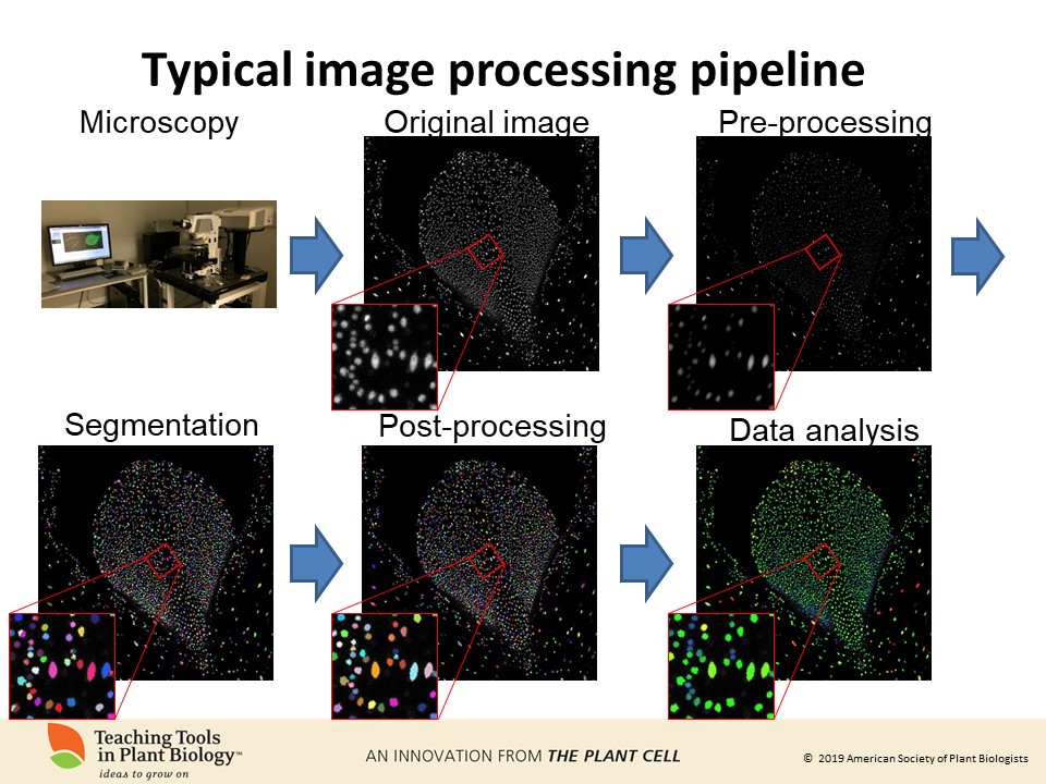
Credit: plantae.org
The Evolution Of Microscopy
Microscopy has transformed our understanding of the microscopic world. From early optical lenses to modern digital imaging, microscopy has evolved significantly. This evolution has enabled scientists to see and study tiny structures in unprecedented detail.
From Optical To Electron Microscopy
The journey of microscopy began with optical microscopes. These use light to magnify small objects. Early optical microscopes revealed cells and simple structures. Despite their limitations, they were revolutionary at the time.
In the 20th century, electron microscopes emerged. These use electron beams instead of light. This allowed scientists to see much smaller objects. Electron microscopes can magnify objects up to 10 million times. They have unveiled the intricate details of viruses and molecules.
Advancements In Imaging Technology
Modern microscopy has seen many technological advances. Digital imaging has enhanced the clarity and precision of microscope images. Confocal microscopy uses lasers to create three-dimensional images. This technique is essential in biological research.
Fluorescence microscopy is another breakthrough. It uses fluorescent dyes to highlight specific structures within cells. This method has made it easier to study complex biological processes.
| Type of Microscopy | Key Features |
|---|---|
| Optical Microscopy | Uses light, magnifies up to 1,000 times |
| Electron Microscopy | Uses electron beams, magnifies up to 10 million times |
| Confocal Microscopy | Uses lasers, creates 3D images |
| Fluorescence Microscopy | Uses fluorescent dyes, highlights specific structures |
Advancements in software have also played a role. Microscopy image processing tools can now enhance and analyze images. These tools help scientists extract more information from their observations.
- Image enhancement
- Noise reduction
- 3D reconstruction
These innovations have made microscopy more powerful and accessible. They continue to push the boundaries of what we can see and understand.
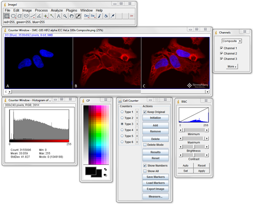
Credit: www.stressmarq.com
Fundamentals Of Microscopy Image Processing
Microscopy image processing is a crucial aspect of analyzing microscopic samples. It helps scientists enhance and interpret images. This process involves several techniques to improve image quality. Understanding these techniques is vital for accurate results.
Pixel Basics And Image Resolution
Every digital image is made up of tiny dots called pixels. The number of pixels in an image determines its resolution. Higher resolution means more detail in the image.
In microscopy, resolution is critical. It allows for clearer and more detailed images. For example, a 1024×768 image has 1024 pixels in width and 768 pixels in height. More pixels provide better clarity and detail.
Contrast Enhancement Techniques
Contrast enhancement makes features in an image stand out. It’s important for highlighting specific structures in microscopy images. Several techniques are used to enhance contrast:
- Histogram Equalization: This technique spreads out the most frequent intensity values. It improves the contrast of the image.
- Adaptive Histogram Equalization: Similar to histogram equalization but more localized. It adjusts contrast in small regions of the image.
- Logarithmic Transformations: This method applies a logarithm to pixel values. It enhances darker regions more than lighter ones.
Using these techniques helps in better visualization of microscopic samples. It allows scientists to observe details that are otherwise hard to see.
Types Of Microscopy Image Processing Tools
Microscopy image processing tools play a crucial role in scientific research. These tools help researchers analyze and interpret microscopic images. Understanding the different types of these tools can greatly enhance research efficiency.
Open-source Vs Proprietary Solutions
Microscopy image processing tools can be categorized into two types: open-source and proprietary solutions.
Open-source tools are free to use and modify. They are often developed by the scientific community. Examples include Fiji and ImageJ. These tools are flexible and customizable. However, they may require more technical expertise to use.
Proprietary solutions are developed by commercial companies. They usually offer more polished user interfaces and dedicated customer support. Examples include Amira and Imaris. These tools often come at a cost. They are generally easier to use but less flexible.
Specialized Software For Different Microscopy Types
Different types of microscopy require specialized software. These tools are designed to handle specific imaging techniques.
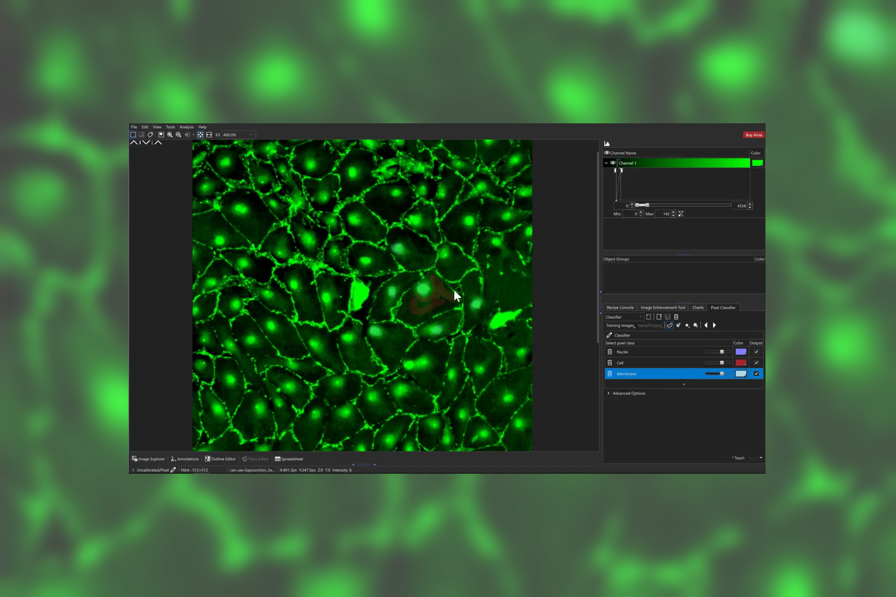
- Confocal Microscopy: Tools like Leica LAS X and Zeiss Zen are tailored for confocal microscopy. They offer features like 3D reconstruction and deconvolution.
- Electron Microscopy: Software such as DigitalMicrograph and Gatan Microscopy Suite are designed for electron microscopy. They provide advanced image analysis and processing capabilities.
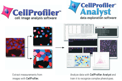
- Fluorescence Microscopy: Tools like CellProfiler and Metamorph are specialized for fluorescence microscopy. They offer functionalities like cell segmentation and quantification.
Choosing the right tool depends on your specific needs. Both open-source and proprietary solutions have their pros and cons. Specialized software ensures optimal performance for different microscopy types.
Credit: www.leica-microsystems.com
Challenges In Microscopic Imaging
Microscopic imaging is crucial for scientific research and medical diagnostics. It reveals details invisible to the naked eye. Yet, it presents challenges that affect image quality and data accuracy.
Dealing With Noise And Artifacts
Microscopic images often suffer from noise and artifacts. These unwanted elements can obscure important details. Noise can stem from various sources:
- Electronic interference
- Light scattering
- Sample preparation issues
Artifacts may result from staining techniques or imaging equipment flaws. They can distort the true structure of the sample. Effective image processing tools are essential for noise reduction. Methods like filtering and deconvolution can help.
Overcoming Limitations Of Resolution
Resolution limits are another challenge in microscopic imaging. Higher resolution reveals finer details but has its limits. Microscopes have a diffraction limit, restricting the smallest resolvable feature.
To overcome this, advanced techniques like super-resolution microscopy are employed. These methods use:
- Structured illumination
- Stimulated emission depletion
- Single-molecule localization
These approaches push beyond traditional limits. They provide clearer and more detailed images. Continuous advancements in microscopy are crucial for scientific progress.
Improving Image Quality
Microscopy image processing tools are vital for researchers. They help improve the quality of microscopic images. Better image quality leads to clearer results. This section covers key techniques to enhance image quality.
Filtering Techniques
Filtering techniques are essential in microscopy image processing. They help remove noise and enhance important features. There are various types of filters used in microscopy:
- Gaussian Filter: Smooths the image by averaging pixel values.
- Median Filter: Reduces noise by replacing pixels with median values.
- Wiener Filter: Minimizes mean square error in the image.
These filters improve image clarity. They make it easier to identify structures in the sample.
Deconvolution Methods
Deconvolution methods aim to reverse the blurring in images. They enhance the resolution and contrast. Two common deconvolution methods are:
- Iterative Deconvolution: Uses algorithms to refine the image over several steps.
- Non-Iterative Deconvolution: Applies mathematical functions to correct blur in one step.
These methods are crucial for achieving high-quality images. They help researchers see fine details clearly.
| Technique | Purpose |
|---|---|
| Gaussian Filter | Smooths the image |
| Median Filter | Reduces noise |
| Wiener Filter | Minimizes error |
| Iterative Deconvolution | Refines image step-by-step |
| Non-Iterative Deconvolution | Corrects blur in one step |
Quantitative Analysis In Microscopy
Quantitative analysis in microscopy helps scientists get precise data. It transforms visual observations into measurable information. This process uses advanced microscopy image processing tools. These tools provide detailed insights into biological samples.
Measuring Intensity And Density
Measuring intensity and density is crucial in microscopy. Intensity indicates the brightness of a pixel. Density reflects the concentration of particles in a sample.
Researchers use software to measure these parameters. They can compare different samples accurately. This helps in identifying specific biological markers and changes.
Automated Cell Counting And Tracking
Automated cell counting saves time and reduces errors. Traditional methods are slow and prone to mistakes. Automated tools can count thousands of cells in seconds.
Tracking cells is also important. It helps in studying cell movement and behavior. Automated tracking tools provide continuous observation. They generate data for dynamic cellular processes.
Below is a table summarizing the benefits:
| Feature | Benefit |
|---|---|
| Automated Counting | Reduces manual errors |
| Automated Tracking | Provides continuous data |
Applications Of Enhanced Microscopy Images
Enhanced microscopy images have revolutionized scientific research. They provide detailed insights into various fields. This section explores their applications in different domains.
Breakthroughs In Biomedical Research
In biomedical research, enhanced microscopy images offer incredible detail. Researchers can observe cellular processes in real-time. This helps in understanding disease mechanisms better.
For example, cancer cells can be tracked with high precision. This aids in developing targeted treatments. Researchers can also study pathogens at a microscopic level. This helps in creating effective vaccines.
Materials Science And Nanotechnology Insights
In materials science, enhanced microscopy images reveal the atomic structure. Scientists can see how materials behave at the smallest scales. This leads to stronger and more durable materials.
Nanotechnology also benefits greatly from microscopy images. Researchers can design nanoparticles with specific properties. This opens up new possibilities in medicine and electronics.
| Field | Application |
|---|---|
| Biomedical Research | Tracking cancer cells |
| Materials Science | Observing atomic structures |
| Nanotechnology | Designing nanoparticles |
The Future Of Microscopy Image Processing
The future of microscopy image processing is bright. New tools and techniques are emerging. They promise better, faster, and more accurate results. These advancements will change how we view microscopic images.
Artificial Intelligence And Machine Learning
Artificial Intelligence (AI) and Machine Learning (ML) are revolutionizing microscopy. These technologies can analyze images faster than humans. They also identify patterns that are hard to spot. This improves accuracy and saves time.
AI can also learn from data. It gets better at recognizing cells or structures. ML models can predict outcomes based on past images. These tools help researchers make informed decisions quickly.
Here are some benefits of using AI and ML in microscopy:
- Faster image analysis
- Improved accuracy
- Pattern recognition
- Predictive modeling
- Automated workflows
Next-generation Imaging Techniques
Next-generation imaging techniques are emerging. They offer better resolution and clarity. These techniques include super-resolution microscopy, light-sheet microscopy, and cryo-electron microscopy.
| Technique | Benefits |
|---|---|
| Super-Resolution Microscopy | Higher resolution than traditional methods |
| Light-Sheet Microscopy | Faster imaging with less light damage |
| Cryo-Electron Microscopy | High-resolution 3D structures of macromolecules |
These techniques allow scientists to see details they couldn’t before. They also reduce the time needed for imaging. This makes research faster and more efficient.
The future of microscopy image processing is promising. With AI and next-generation techniques, the possibilities are endless.
Frequently Asked Questions
What Is The Image Processing Method In Microscopy?
Image processing in microscopy involves enhancing, analyzing, and visualizing microscopic images using software tools. Techniques include filtering, segmentation, and 3D reconstruction. These methods improve image clarity, highlight features, and extract quantitative data, aiding in accurate scientific analysis.
What Is The Best Software For Sem Image Analysis?
The best software for SEM image analysis includes ImageJ, Fiji, and MATLAB. These tools offer advanced features and user-friendly interfaces.
Which Tool Is Used For Image Analysis?
The tool commonly used for image analysis is Adobe Photoshop. Other popular options include GIMP and MATLAB.
What Is The Free Software For Microscopes?
Free software for microscopes includes ImageJ, Fiji, and Micromanager. These tools offer image analysis, processing, and control.
Conclusion
Microscopy image processing tools enhance research and diagnostics. They offer precision and efficiency, vital for scientific advancements. By leveraging these tools, researchers can achieve clearer, more detailed images. This leads to better insights and breakthroughs. Stay updated with the latest tools to maximize the potential of your microscopy work.